From the microscopic architecture of fungi and insects to high-tech machinery and delicate crystals, the winning entries in the 2026 Nikon Small World Photomicrography Competition have once again pulled back the curtain on astonishing, intricate details that remain entirely invisible to the naked eye. For decades, this prestigious competition has served as a bridge between the worlds of art and science, celebrating the incredible imagery captured by researchers, technicians, and hobbyists who peer through microscope lenses to document the hidden mechanics of our universe.
This year’s highest honor—the top prize—was awarded to Ole Bielfeldt, a student and experimental photographer based in Cologne, Germany. To secure the victory
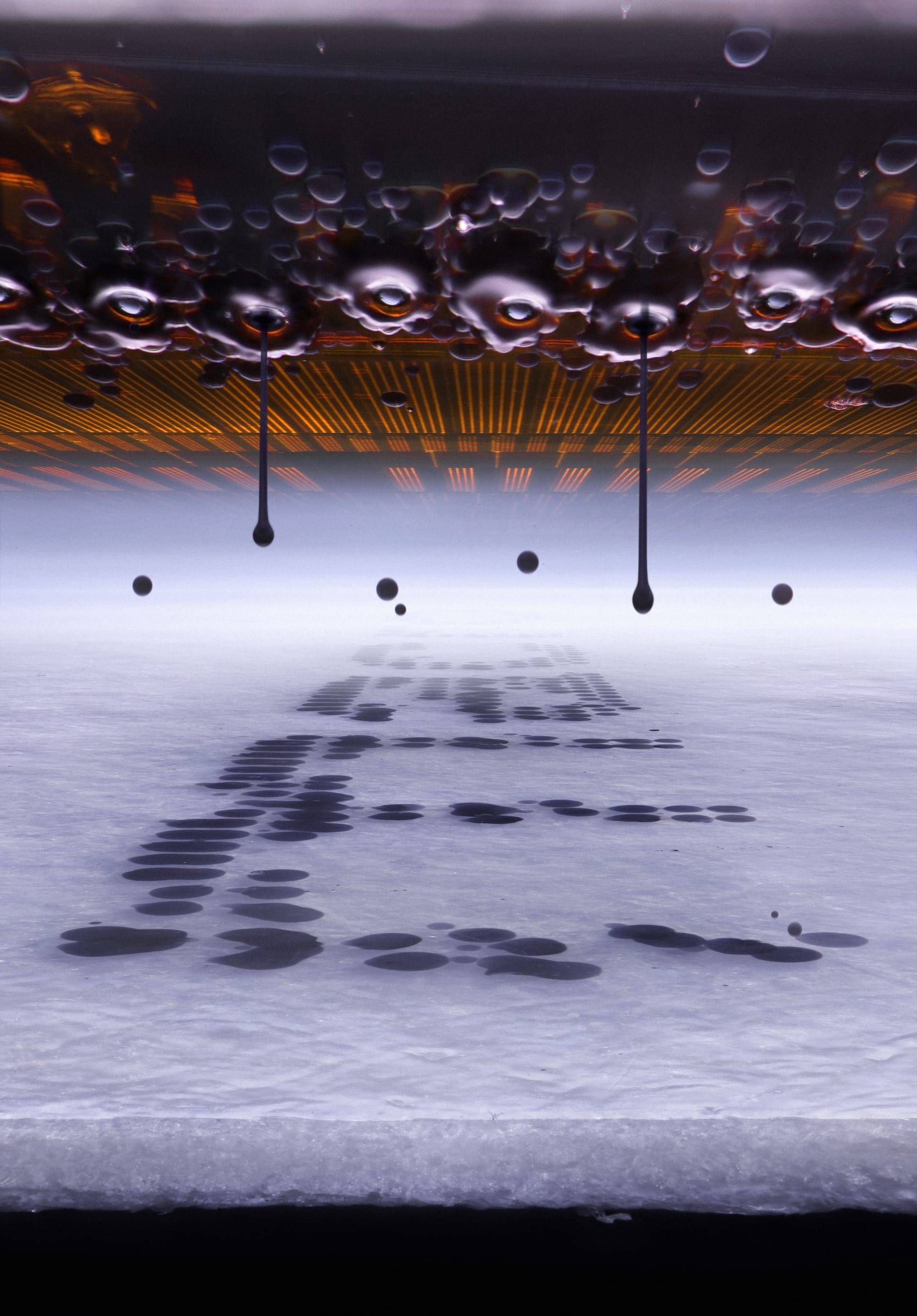
, Bielfeldt utilized a specialized, hand-built imaging rig to capture an extraordinary, hyper-detailed up-close view of microscopic droplets inside an inkjet printer as ink is actively deposited onto a sheet of paper. The resulting image showcases a precise intersection of modern office technology and micro-scale fluid dynamics, freezing a fraction of a second where liquid meets media in a vibrant display of engineering and physics.
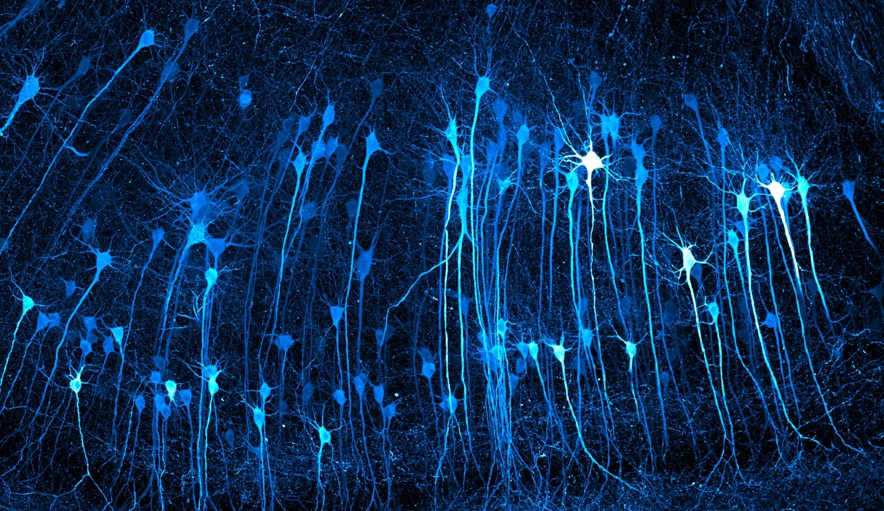
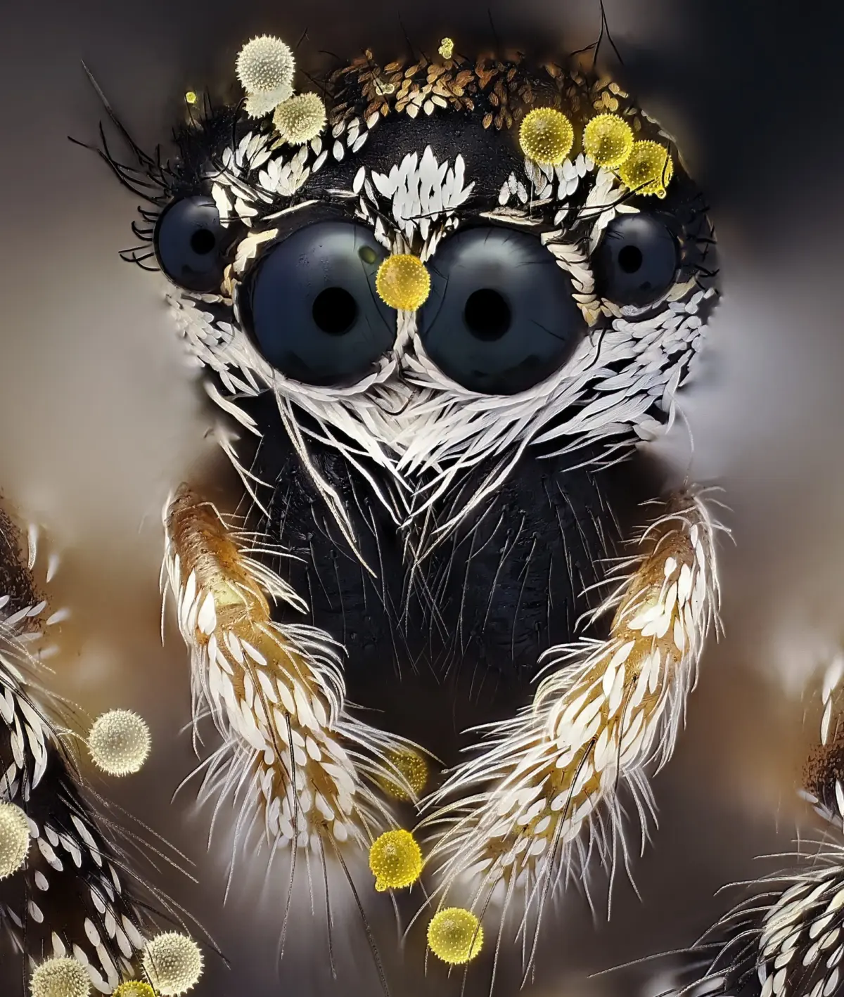
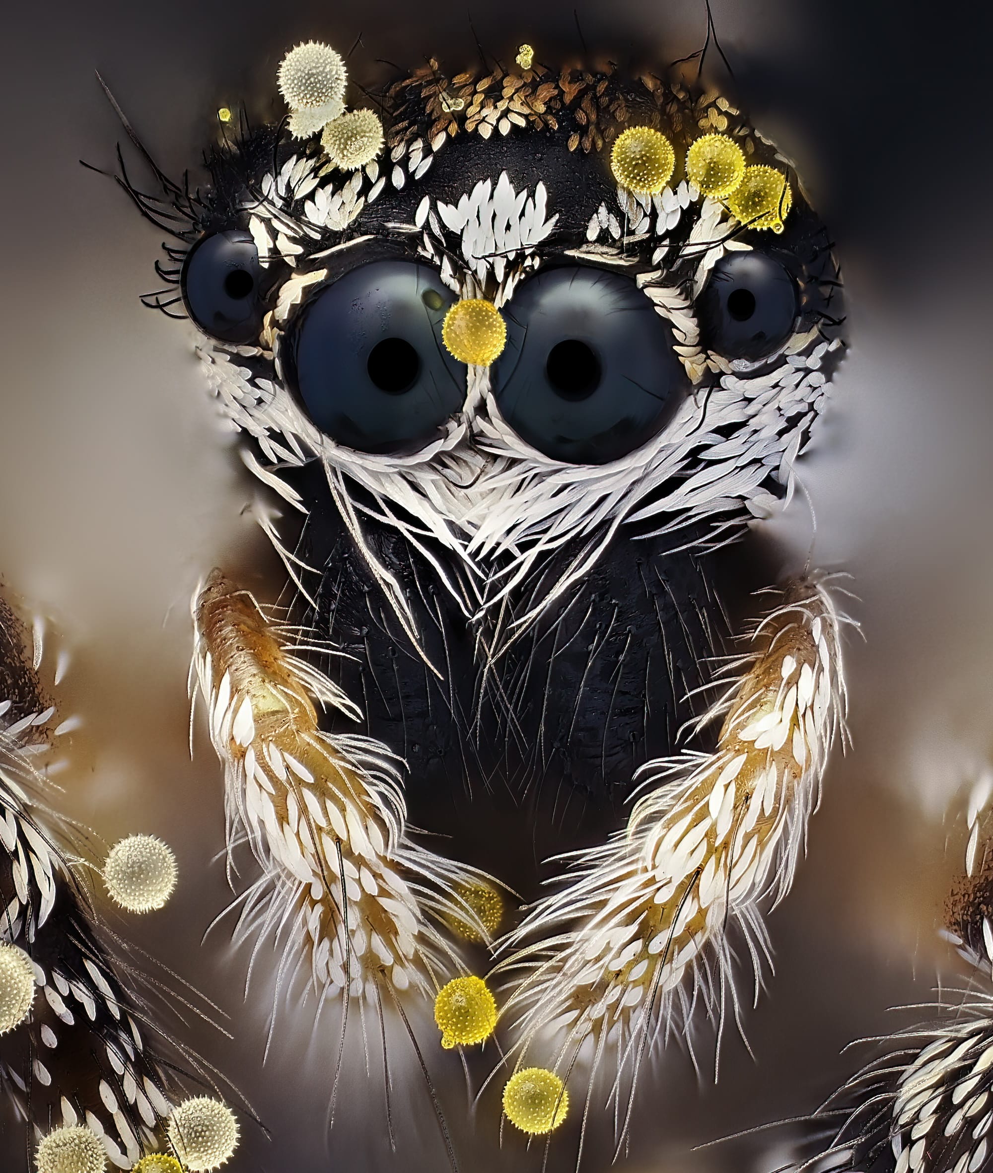
While Bielfeldt claimed the ultimate prize, the broader selection of top-tier winners and runners-up spans a vast array of scientific disciplines and natural wonders. Among the standout entries is a captivating portrait captured by John-Oliver Dum, which highlights a jumping spider dusted with golden grains of pollen across its body. In another striking display of biological imaging, Kaelin Wulf and Rachel Flannery of the Allen Institute’s Department of Neural Dynamics in Seattle, Washington, earned high praise for their intricate capture of neurons within an expanded mouse brain, illuminating the complex network of the mammalian nervous system in breathtaking clarity.
The competition organizers organized this year’s exceptional pool of winner images into three primary tiers: the Top 20, Honorable Mentions, and Images of Distinction. Science and photography enthusiasts alike can explore these selections directly on the contest’s official website, which hosts a comprehensive gallery of the year’s finest microscopic photography. For those interested in moving imagery, the contest also runs a companion initiative known as the Nikon Small World in Motion video competition, which highlights the dynamic, shifting behaviors of microscopic subjects over time.
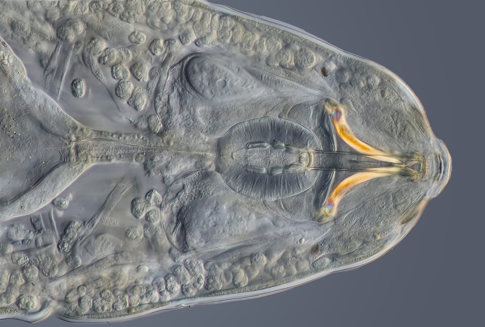
A closer look at the honored works reveals a diverse cross-section of life and materials captured across the globe. Among the recognized pieces is an image by Charles Krebs of Issaquah, Washington, categorized as an Image of Distinction, which features a tardigrade—commonly known as a "water bear"—with its piercing stylets and pharynx clearly exposed, offering a rare look at the resilient micro-animal’s feeding apparatus.
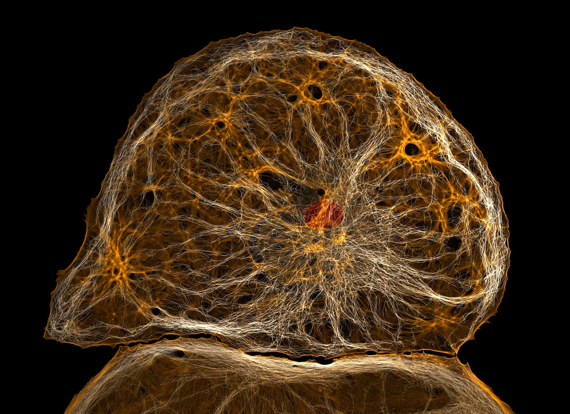
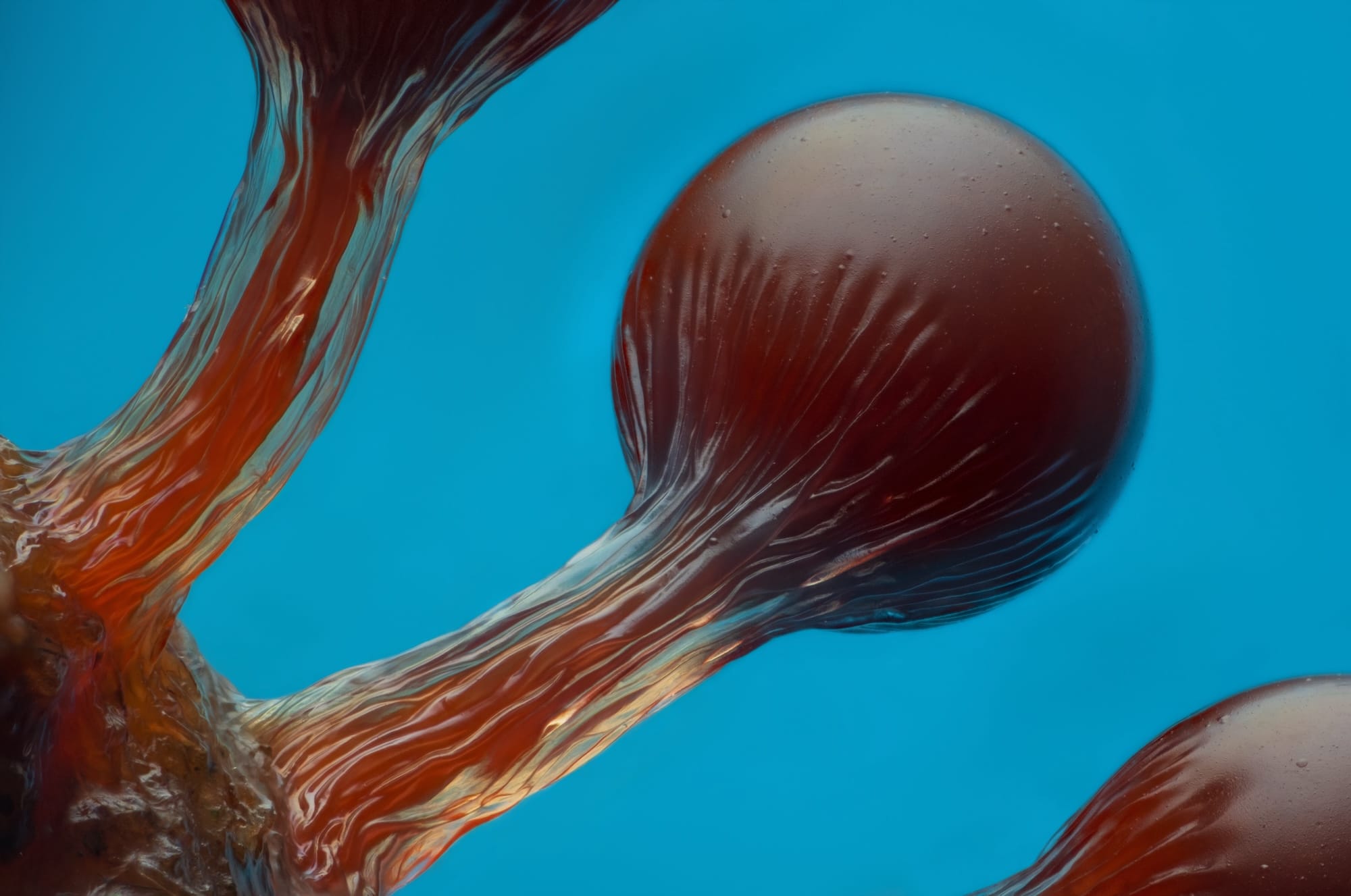
In the sixth-place position, Eric Epstein of Brooklyn, New York, submitted a striking view of Hemitrichia slime mold spore structures, highlighting the delicate, maroon-colored fruiting bodies of the organism against a contrasting blue background. Meanwhile, Dr. Francisco Lázaro-Diéguez of the Albert Einstein College of Medicine in the Bronx, New York, captured the ninth-place entry, focusing on a dedifferentiated liver endothelial cell that reveals the complex cellular architecture underlying human and animal biology.
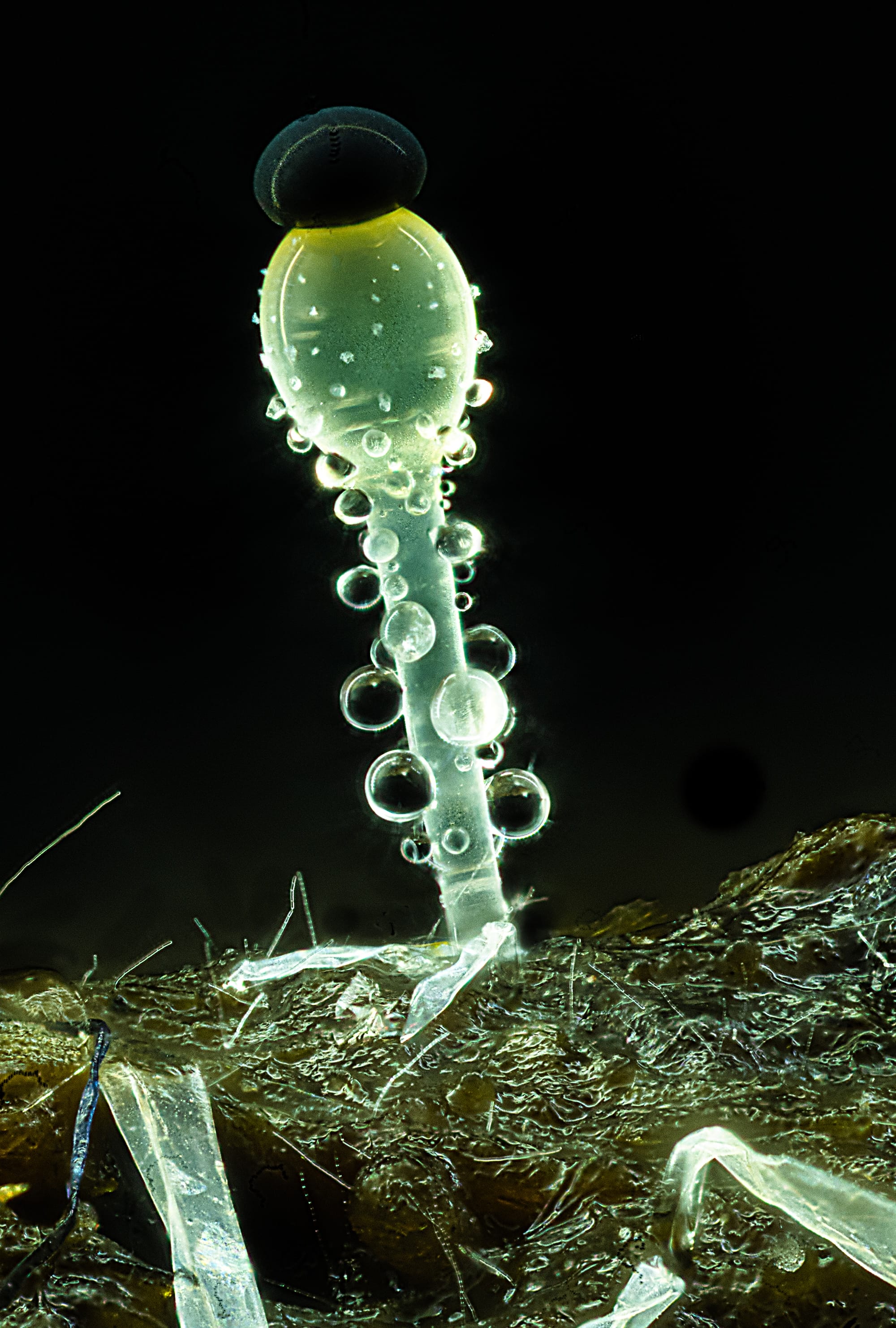
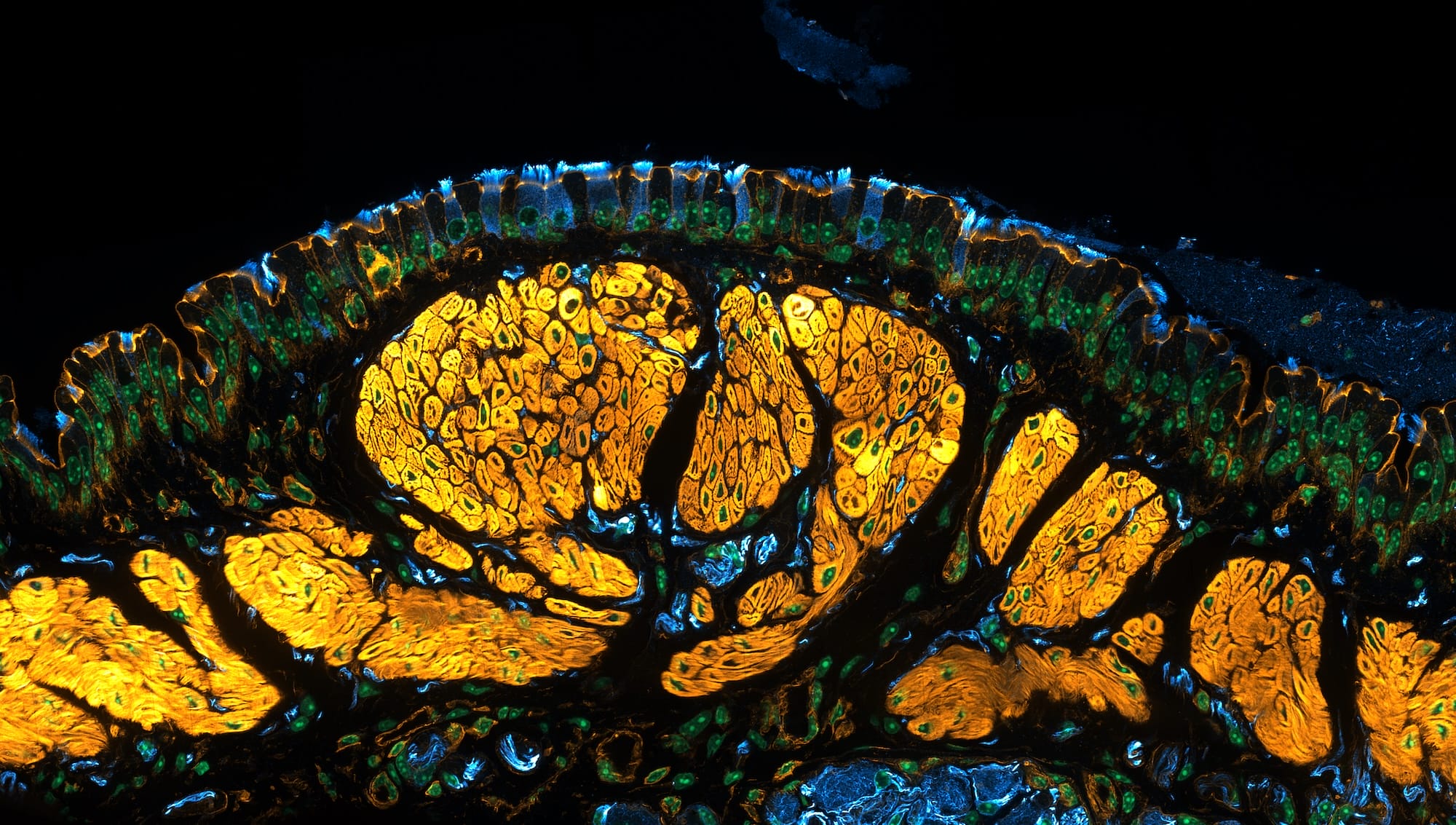
The natural world’s smaller kingdoms are further represented by Mike Crutchley of Pembrokeshire, Southwest Wales, who secured the 14th-place spot with a macro-microscopic composition of Pilobolus fungi thriving on sheep dung. Not far behind in 15th place, Dr. Julien Resseguier of the University of Oslo’s Department of Biosciences in Norway presented a detailed look inside a mouse trachea. The image vividly demonstrates tiny, hair-like cells responsible for moving mucus, alongside surrounding blue-tinted nerves linked to yellow-highlighted muscular tissue.
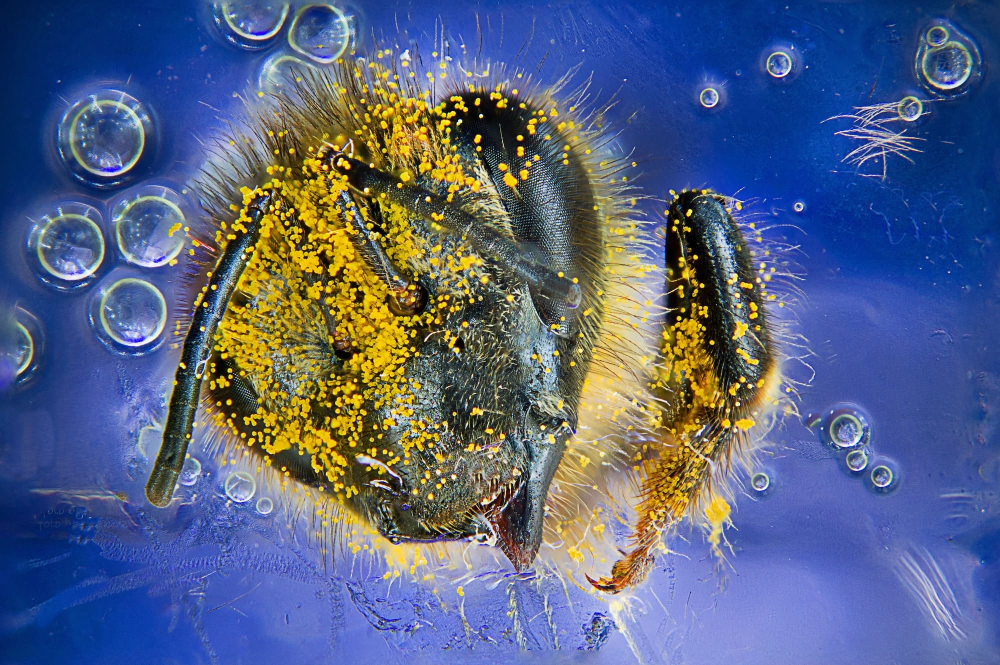
Other notable entries earning Images of Distinction include a detailed portrait of a Western honey bee (Apis mellifera) heavily laden with pollen, captured by Giuseppe Franchina of Messina, Italy, and a remarkably symmetrical geometric study of a single snowflake viewed under a microscope by Sergey Kichigin of Vologda, Russia. In the Honorable Mention category, Jan Martinek of the Czech Academy of Sciences’ Institute of Experimental Botany in Prague, Czech Republic, captured a common daisy flower head (Bellis perennis) glowing vibrantly under its own natural autofluorescence.
These works, along with the complete collection of 2026 entries, continue to demonstrate how advanced optics and artistic vision can transform ordinary scientific samples into profound visual art.